TL;DR
A Griffith University research team used advanced, multimodal MRI to compare people with Long COVID, people who considered themselves fully recovered, and never-infected controls, finding measurable changes in brain tissue and neurochemical profiles. Altered brain signals and tissue structure were linked to symptom severity in Long COVID and were present even in some people without ongoing symptoms.
What happened
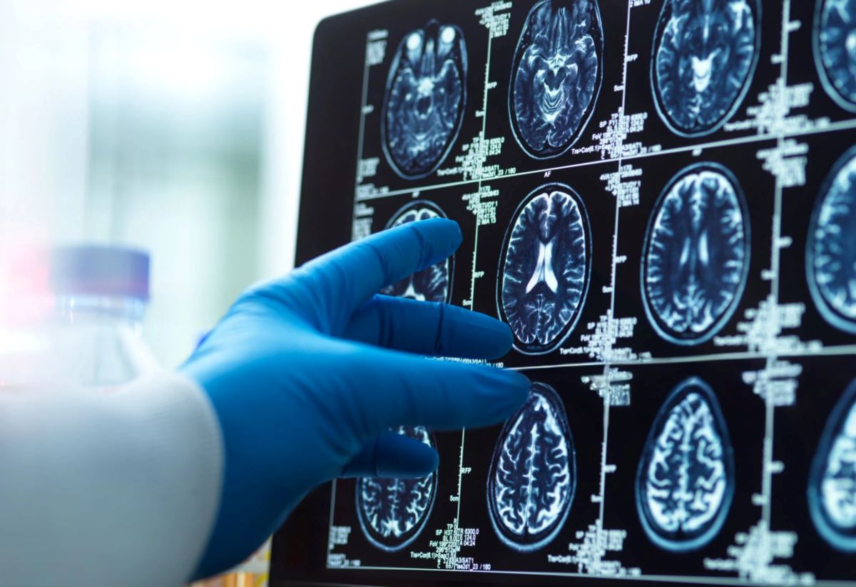
Researchers at Griffith University’s National Centre for Neuroimmunology and Emerging Disease applied a suite of MRI methods to examine both grey and white matter regions involved in memory and cognition. The study identified clear differences in brain neurochemistry, MRI signal intensity and microstructure when comparing people with Long COVID, individuals who considered themselves recovered, and those never infected. Notably, changes were detected not only in participants reporting persistent symptoms but also in some who reported full recovery. The team reported an association between altered brain tissue and the severity of Long COVID symptoms. The work was funded by ME Research UK and the Stafford Fox Medical Research Foundation and published as ‘Altered Brain Tissue Microstructure and Neurochemical Profiles in Long COVID and Recovered COVID-19 Individuals: A multimodal MRI Study’ in the journal Brain, Behavior, and Immunity – Health.
Why it matters
- Provides evidence that SARS-CoV-2 infection can leave measurable changes in the central nervous system beyond the acute phase.
- Helps account for cognitive complaints such as memory loss and difficulty concentrating reported after infection.
- Suggests that even people who feel fully recovered may have subtle brain changes detectable by advanced imaging.
- Supports the case for continued research and clinical monitoring of neurological outcomes after COVID-19 infection.
Key facts
- Study conducted by Griffith University’s National Centre for Neuroimmunology and Emerging Disease (NCNED).
- Researchers used multimodal MRI to examine both grey and white matter regions tied to memory and cognition.
- Findings included alterations in brain neurochemicals, MRI signal intensity, and tissue microstructure.
- Changes were observed in participants with Long COVID and in some who considered themselves fully recovered.
- Altered brain tissue measures were associated with symptom severity among individuals with Long COVID.
- The study paper was published in Brain, Behavior, and Immunity – Health.
- Research funding came from ME Research UK and the Stafford Fox Medical Research Foundation.
- Authors highlighted the potential of advanced imaging to reveal lasting effects of COVID-19 on brain health.
What to watch next
- Whether independent groups replicate these MRI findings in larger and diverse cohorts: not confirmed in the source.
- Longitudinal studies tracking whether the observed brain changes persist, progress or recover over time: not confirmed in the source.
- Research clarifying mechanisms linking SARS-CoV-2 infection to the specific neurochemical and microstructural changes seen on MRI: not confirmed in the source.
Quick glossary
- Multimodal MRI: The use of multiple magnetic resonance imaging techniques that capture different aspects of brain structure, chemistry and function to provide a comprehensive view.
- Grey matter: Brain tissue rich in neuronal cell bodies involved in processing information, cognition and memory.
- White matter: Brain tissue composed mainly of nerve fibres (axons) that connect different brain regions and support communication between them.
- Long COVID: A term used for a range of symptoms that continue or develop after the acute phase of a SARS-CoV-2 infection.
- Neurochemical profile: The concentrations and balance of chemicals in the brain that influence neuronal function and signalling.
Reader FAQ
Does the study show COVID-19 causes permanent brain damage?
The study reports measurable alterations in brain tissue and chemistry after infection, but permanence of these changes is not confirmed in the source.
Were changes seen only in people with ongoing symptoms?
No. The study found alterations in both people with Long COVID and in some participants who considered themselves fully recovered.
Where was the study published and who funded it?
The paper appeared in Brain, Behavior, and Immunity – Health and was funded by ME Research UK and the Stafford Fox Medical Research Foundation.
Can routine clinical MRI detect these differences?
Not confirmed in the source.
COVID-19 does not just affect the respiratory system, but also significantly alters the brain in people who have fully recovered from the infectious disease, highlighting the long-term neurological impact of…
Sources
- Covid-19 leaves a lasting mark on the human brain
- COVID-19 Leaves Lasting Changes in the Brain, Even …
- Brain MRI findings in patients with post COVID-19 condition
- Distinct brain alterations and neurodegenerative processes …
Related posts
- Go.sum is not a lockfile — stop parsing it to analyze dependency graphs
- Lean 4 proof argues SSOT needs definition-time hooks and introspection
- Go authors: stop treating go.sum as a lockfile — use go.mod instead